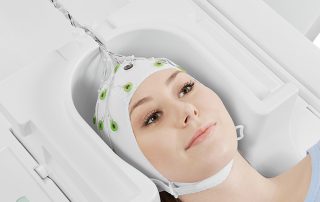
Exciting news for our EEG-fMRI customers: Update of MRI sequence conditions
We are excited to announce some news that will bring our conditions for use in line with current trends in fMRI. We have updated the MRI sequence conditions for our BrainAmp MR amplifiers and BrainCap MR.