How to decide which EEG amplifier best fits your research
by Sara Pizzamiglio, PhD, Eduardo Bellomo, PhD and Laura Leuchs, PhD
Scientific Consultants (Brain Products)
 Deciding which EEG amplifier is the best for your research work can be challenging. For some applications, specific requirements may hint at a well-defined solution. When no specific needs are to be considered, the choice may instead be guided by other technological and practical aspects.
In this article we first highlight the main features of all Brain Products EEG amplifiers, from laboratory-based to mobile and wearable. We then offer some guidelines on how to decide on an EEG amplifier based on factors like; the technical features of the devices, their usability features, and the application for which it will be used.
Introduction
When setting up a new lab or starting a new line of investigations, you may find yourself wondering which is the right EEG system for your work. Will you be recording data in a protected and shielded environment? Will you take your experiment out of the lab into the real world? Or will you use EEG simultaneously with other stimulation or measurement techniques?
Based on your application, you may prefer or even require a specific system; if not, you may still have your own preferences. With this article we would like to present to you all the EEG amplifiers that Brain Products currently offers and share some guidelines on how to choose the best system for your work.
1. What is an EEG amplifier?
As you know, surface Electroencephalography (EEG) is the non-invasive measurement of the electrical activity of the human brain. When many neurons activate synchronously in the brain, the total potential resulting from the sum of their simultaneous postsynaptic potentials is detected at the level of the scalp (i.e., brain tissue, skull, muscles, skin). To measure this signal you need surface electrodes to register the brain electrical activity and an operational amplifier to amplify, convert, and transmit the signal to the recording computer for further processing and saving.
The work of an EEG amplifier is quite simple to understand and can be summarized in just a few steps:
Tips:
- Watch the recording of our Brain Products Academy workshop session on EEG Hardware if you are new to the EEG world and would like to know more on how an amplifier works and how to ensure high quality data.
- Read this article to know more on important factors that you should consider when choosing your online and offline reference.
2. Brain Products amplifiers: an overview
All our amplifiers* work with BrainVision Recorder, our comprehensive software for data recording. It allows you to define all your recording settings (e.g., sampling frequency, total number of channels and layout, digital ports, etc.), to prepare the cap via the live impedance mode, to monitor the data online, and ultimately save them in your preferred PC location. It also allows you to stream your data to any third-party application (e.g., MATLAB®, C, C++, etc.) thanks to the Remote Data Access (RDA) option.
Let us now have a look at each one of our systems and highlight their stand out characteristics. You can also find a complete overview of the technical features in the Amplifier Comparison Table available for download.
Tips:
- If data streams from many different sources need to be collected in a synchronized fashion, as an alternative to the RDA you can use LabStreamingLayer (LSL, an open-source solution from the SCCN, Kothe et al., 2014). Read more on how you can achieve this on our dedicated BCI+ blog post.
- If you are planning a multicenter study where different setups may be involved, make sure to read our tips for comparability across sites in this article.
* except for the CGX Mobile series (see the dedicated section below)
2.1. Laboratory-based solutions
2.1.1. Our state-of-the-art: the actiCHamp Plus
Born as an evolution of its predecessor (i.e., the actiCHamp), the actiCHamp Plus is our state-of-the-art stationary solution. The Plus feature of this latest generation lies in the possibility to record data from both active or passive electrode technologies (i.e., the actiCHamp was compatible only with active electrodes). Powered by a rechargeable Lithium-ion (Li-ion) battery called the PowerUnit (Figure 1 bottom), the actiCHamp Plus comes as a scalable single-base unit (Figure 1, upper panel of top unit) that can be equipped with up to five 32 channel EEG modules (i.e., maximum 160 channels). The front panel has 8 Auxiliary (AUX) inputs for the recording of peripheral physiological signals simultaneously with the EEG. Recorded data is transmitted via USB to the recording computer, where they are eventually saved.
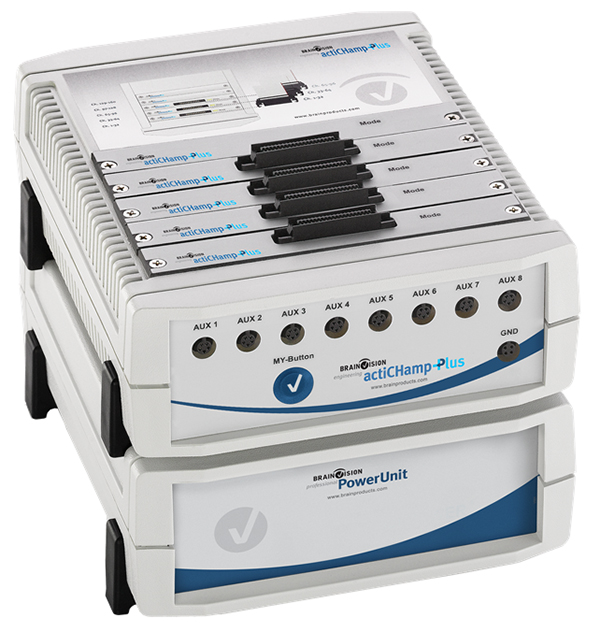
Figure 1: 128-channel actiCHamp Plus amplifier with PowerUnit (rechargeable battery).
2.1.2. Our classics: the BrainAmp family
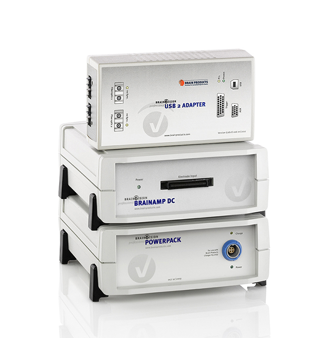
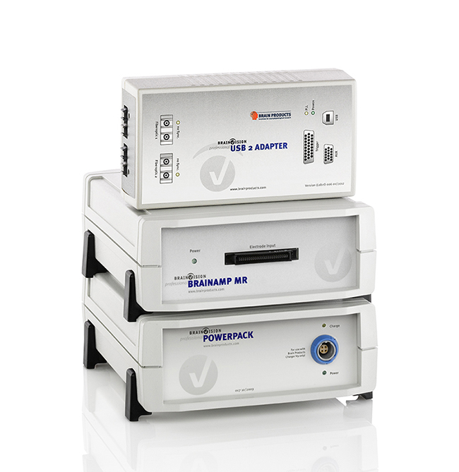
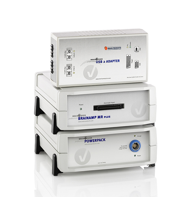
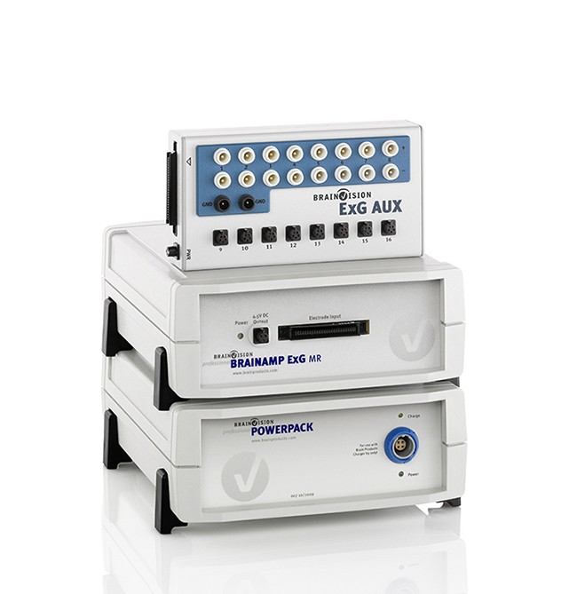
Figure 2: The BrainAmp family. BrainAmp Standard and DC for EEG recordings, and BrainAmp Exg for peripheral physiology in the lab environment.
BrainAmp MR and MR plus for EEG recordings and BrainAmp ExG MR for peripheral physiology in the MR environment.
Since its inception as one of the first amplifier families launched by Brain Products, the BrainAmp systems rapidly became the gold-standard in EEG laboratories and contributed to thousands (and counting) of publications. The family includes 4 unipolar EEG and 2 bipolar ExG amplifiers, which differ in both their technical features (see the Amplifier Comparison Table for details) and, most importantly, in their compatibility with the MR environment.
These EEG systems come in 32-channel units powered by a lead rechargeable battery, the PowerPack. A maximum of 256 EEG channels can be recorded in laboratory settings (i.e., max. 8 units), whilst data from up to 128 EEG channels can be acquired within the MR (i.e., max. 4 units) environment. Originally, the BrainAmp family of amplifiers was designed to work only with passive electrode technology, but thanks to specific adapters it is now also possible to combine them with active electrodes (i.e., via the actiCAP ControlBox II). If you want to record peripheral physiological signals, you can use the optional ExG amplifier units together with the ExG AUX Box (8 touchproof bipolar and 8 AUX inputs). Data is recorded and transmitted via double fiber optic cables to the BrainAmp USB 2 Adapter (BUA), an intermediate interface that synchronizes data streams from all connected BrainAmp amplifiers for the subsequent transmission via USB to the recording computer.
Tips:
- One battery can power two BrainAmp units at the same time and it is possible to combine EEG and ExG acquisition systems; but be careful when doing so in the MR environment. Read our safety tips for EEG-fMRI measurements in this article and watch the related webinar.
- If you work in the EEG-fMRI field and would like to add Carbon Wire Loops to your BrainCap MR for even more accurate MR-related artifacts removal, you may be interested in the BrainAmp ExG MR 8 ch version (see this article for more information).
2.2. Mobile and wearable solutions
2.2.1. Compact, light, and wireless: the LiveAmp
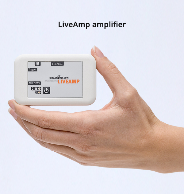
The LiveAmp was designed for applications that require reliable wireless recordings. Data can be transmitted via a wireless protocol to the recording computer and/or saved directly on an exchangeable memory card. Available in various upgradable channel count versions (i.e., 8, 16, 32 and 64), the LiveAmp is powered by an internal Li-ion rechargeable battery, which lasts approximately 4 hours (depending on the recording modality and electrode technology, see Amplifier Comparison Table).
The LiveAmp is compatible with both passive and active electrode technologies, and even has a built-in 3D Accelerometer for the recording of movement acceleration in the three dimensional space.
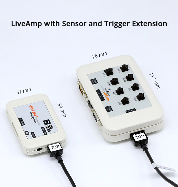
Furthermore, if you need to record peripheral physiological signals or to mark multiple events with more complex triggers (i.e., up to 8-bit), you can extend the system by connecting the Sensor and Trigger Extension (STE).
Tips:
- If longer recording times are required, you can connect an external power bank directly to the amplifier and run your experiment for as long as you need to. If you are using the STE, an external power bank is always required.
- Our Wireless Trigger gives you the chance to set up fully mobile experiments and still receive triggers reliably. Connect the Receiver unit to the STE and the Transmitter to our Trigger Box for optimal data synchronization.
2.2.2. All-in-one and one-size-fits-all: the CGX Quick systems
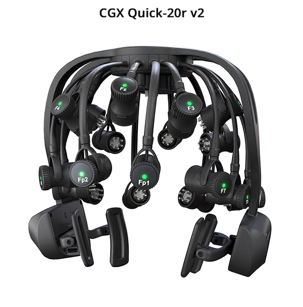
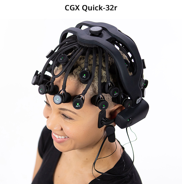
The new generation of Quick systems are the state-of-the-art active dry, wireless EEG headsets, born from the strong partnership between Brain Products and CGX. They are an all-in-one solution, whereby the integrated amplifier is now located on the left ear pad and the rechargeable batteries on the right ear pad for a more balanced headset.
The new Flex Sensors design consists of smaller tips and electrode pods together with stronger pod-hinges, which together ensure higher comfort, stability, and data quality. The design still follows the one-size-fits-all approach, whereby one headset fits heads sizes in a range from 52 cm to 62 cm (i.e., adolescents through adults). Moreover, LED impedance indicators (a patented technology by Brain Products) have been embedded into each Sensor Pod for an even better user experience and faster preparation time. The Quick systems are available in 20 and 32 channel versions.
You can combine your EEG recording with other modalities by using the AIM Physiological Monitor, a wireless lightweight amplifier equipped with 6 bipolar and 2 AUX inputs for measuring ExG (i.e., EMG, ECG), GSR, temperature, respiration, heart rate (HR), pulse and SpO2. Moreover, hardware wireless triggers up to 16-bits can be transmitted to any CGX device via the Wireless StimTrigger, which accepts incoming signals from many different sources (e.g., light and audio sensor, microphone, USB, TTL, etc.).
Both the Quick systems and the AIM work seamlessly with BrainVision Recorder for CGX as well as with CGX Acquisition.
Notes:
- Two separate computers running separate instances of BrainVision Recorder for CGX or CGX Acquisition are required to record data from the EEG and physiological amplifiers, respectively. When using BrainVision Recorder, two separate USB dongles (therefore, two separate licenses) are required, one plugged into each computer.
- The Quick systems are powered by two AA-type supplied rechargeable batteries which, when new and fully charged, have a battery life of 8 hours. The AIM Physiological Monitor is powered by a built-in rechargeable Li-on battery which, when new and fully charged, has a life of 5 hours.
2.2.3. High-density and wireless: the CGX Mobile
The CGX Mobile systems are a unique high-density wireless solution, available as 72 (figure 3) or 128 active gel-based channel versions. The system is fully mobile and compact: it comes with a harness that is equipped with an electrode bundle Breakout Box. The small amplifier, powered by a rechargeable Li-ion battery, can be attached to the Breakout Box. The lead wires are bundled together into a single cable tree, which exits from the same Breakout Box.
As with the Quick systems, you can combine the Mobile amplifiers with the AIM Physiological Monitor and receive wireless triggers via the Wireless StimTrigger.
Notes:
- The CGX Mobile systems work only with the CGX Acquisition software.
- Both the CGX Quick and Mobile systems have a fixed number of electrodes which cannot be changed.

Figure 3: CGX Mobile 72
3. How to choose the right amplifier for your application?
You may appreciate that deciding between two amplifiers can be tricky sometimes. In this section we would like to give you some guidelines on how to approach the choice of the EEG system that fits with your goals. Eventually, you may find yourself looking for a compromise between application-specific needs, desired signal content requirements, your current budget, and future possibilities. The table below summarizes some of the aspects that we will cover in the next section and provides an overview of the qualitative features of each one of our EEG amplifier solutions.
Table: Brain Products EEG amplifiers’ qualitative features overview (currently available systems for purchase)
| BrainAmp Standard |
BrainAmp DC |
BrainAmp MR |
BrainAmp MR plus |
actiCHamp Plus |
LiveAmp | CGX Quick |
CGX Mobile |
|
|---|---|---|---|---|---|---|---|---|
| Time resolution (Sampling Rate, SR) | good switchable SR up to 5 kHz |
good switchable SR up to 5 kHz |
good switchable SR up to 5 kHz |
good switchable SR up to 5 kHz |
excellent switchable SR up to 100 kHz |
good switchable SR up to 1 kHz |
fair fixed SR of 500 Hz |
fair fixed SR of 500 Hz |
| Measurement range and amplitude resolution | fair fixed range of ±3.28 mV at 0.1 µV/bit |
good switchable: ±3.28 mV at 0.1 µV/bit; ±16.384 mV at 0.5 µV/bit; ±327.68 mV at 10 µV/bit** |
good fixed range of ±16.384 mV at 0.5 µV/bit |
good switchable: ±3.28 mV at 0.1 µV/bit; ±16.384 mV at 0.5 µV/bit; ±327.68 mV at 10 µV/bit** |
excellent fixed range of ±409.6 mV at 0.0487 µV/bit |
excellent fixed range of ±341.6 mV at 40.7 nV/bit |
excellent fixed range of ±833 mV at 0.1 µV/bit |
excellent fixed range of ±833 mV at 0.9 µV/bit |
| Scalability (max. channel number) |
excellent up to 256 ch |
excellent up to 256 ch |
excellent up to 128 ch inside MR, up to 256 ch in lab environment |
excellent up to 128 ch inside MR, up to 256 ch in lab environment |
excellent up to 160 ch |
good up to 64 ch |
n/a fixed channel number (20 or 32) |
n/a fixed channel number (72 or 128) |
| Setup complexity | complex multiple amp units and batteries required |
complex multiple amp units and batteries required |
complex multiple amp units and batteries required |
complex multiple amp units and batteries required |
very simple one amp unit with multiple modules and one battery |
simple multiple but small units |
very simple all-in-one and small units |
simple multiple but small units |
| Versatility | good compatible with passive gel- and sponge-based electrodes; requires actiCAP ControlBox II for active gel-based and dry electrodes |
good compatible with passive gel- and sponge-based electrodes; requires actiCAP ControlBox II for active gel-based and dry electrodes |
good compatible with passive gel- and sponge-based electrodes for EEG-fMRI; outside the MR compatible also with active gel-based and dry electrodes (requires actiCAP ControlBox II) |
good compatible with passive gel- and sponge-based electrodes for EEG-fMRI; outside the MR compatible also with active gel-based and dry electrodes (requires actiCAP ControlBox II) |
excellent compatible with active gel-based and dry, as well as with passive sponge-based electrodes, requires EIB64DUO for passive gel-based electrodes |
excellent compatible with active gel-based and dry, as well as with passive gel- and sponge-based electrodes |
n/a active dry electrodes only |
n/a active gel-based electrodes only |
| Real-time accessibility | fair transmission in tenths of ms via LSL |
fair transmission in tenths of ms via LSL |
fair transmission in tenths of ms via LSL |
fair transmission in tenths of ms via LSL |
excellent transmission in <1.5 ms via TurboLink |
good transmission in few ms via LSL |
good transmission in few ms via LSL |
good transmission in few ms via LSL |
| Simultaneous TMS | poor | excellent | poor | excellent | good | fair | poor | fair |
| Simultaneous MEG | yes | yes | yes (the best option) |
yes (the best option) |
under evaluation |
no | no | no |
| Simultaneous MR | no | no | yes (MR Conditional) |
yes (MR Conditional) |
no | no | no | no |
| Mobile / Wearable | no | no | no | no | no | yes | yes | yes |
**Please note that for all BrainAmp family amplifiers the measurement range of ±327.68 mV at 10 µV/bit does not represent a physiological measurement range/resolution, but can be used for troubleshooting or e.g. trigger signals.
3.1. Technical features
When planning your investigations, you should always first make sure you have a system that can actually provide you with the information you are looking for. The conversion of the time-wise continuous EEG signal into discrete digital data points is a crucial part of an amplifier and ADC are characterized by:
3.1.1. Time resolution & Sampling Rate (SR)
These define the regular time intervals at which data points of the continuous EEG signal are acquired. The sampling rate determines how many data points per second you can record and influences the frequency content available in the recorded EEG signal. Insufficient time resolution leads to the loss of important information, or to phenomena like the aliasing effect, whereby two different signals cannot be properly distinguished from each other when sampled. Aliasing causes the appearance of fictional content in the recorded data that was not part of the original signal. To avoid this, the recording software (in our case, BrainVision Recorder) applies an antialiasing filter at ¼ of the sampling rate. Insufficient sampling can also lead to the loss of important information. According to the Nyquist theorem, a sampling rate of at least twice the value of the frequency content of interest should be used to reliably capture it (Nyquist, 1928), and it is even further suggested to acquire data at a rate at least 5 – 10 times higher than the signal of interest (Weiergräber et al., 2016). For example, to properly record Event Related Potentials within a frequency range between 1 Hz and 50 Hz, a sampling rate >250 – 500 Hz should be used.
Based on the frequency content of your EEG signal, you might want a system that offers sufficiently/very high or switchable temporal resolution. In these cases, the actiCHamp Plus is the best option for you since it can record data with different SRs up to 100 kHz (depending on the total number of channels). For example, a sampling rate of 25 kHz will allow you to minimize the effects of a TMS stimuli on the recorded EEG data (i.e., minimal ringing artefacts), thus improving the overall quality of the recording. High sampling rates of 25 kHz and 50 kHz are also recommended nowadays when investigating Auditory Evoked Potentials in order to acquire more accurate signals and clearer artifacts of cochlear implants (Attina et al., 2017). In addition, if high quality and unfiltered audio signals are needed simultaneously to the EEG, you may record up to ~16 kHz signals via one of the AUX ports at 100 kHz sampling rate.
If your research does not require a very high sampling rate, you might still choose the actiCHamp Plus for the other advantages it offers (see below). If you are unsure about the frequency content you need, having the possibility to choose high sampling rates will leave many doors open to you.
3.1.2. Amplitude resolution & bit depth
These define the regular voltage intervals at which the continuous EEG signal’s amplitude is digitized. The number of available bits of an amplifier represents the bit depth and determines how many possible digital output values a signal can have (i.e., 2total_bit). The more possible output values are available, the more fine-grained the amplifier can differentiate between different voltage steps. Together, the bit depth and the amplitude resolution determine the amplifier measurement range, that is the maximum and minimum voltage values that can be recorded.
Insufficient measurement range can prevent the recording of a signal whose values exceed the available range (i.e., clipping effect). Based on the strength of your EEG signal of interest, you might want a system with a regular or a more generous range. For example, if you are working in the EEG-TMS field, you want a system that can record the full signal even during stimulation and the actiCHamp Plus will surely fit your needs well (measurement range of ±409.6 mV at 0.0487 µV/bit). Alternatively, the BrainAmp DC and the BrainAmp MR plus could also be valid options, thanks to the switchable resolution and measurement range features they offer (measurement range of ±3.28 mV at 0.1 µV/bit, ±16.384 mV at 0.5 µV/bit, ±327.68 mV at 10 µV/bit**).
**Please note that for all the BrainAmp family amplifiers the measurement range of ±327.68 mV at 10 µV/bit does not represent a physiological measurement range/resolution, but can be used for troubleshooting or e.g. trigger signals.
3.2. Usability features
If you are starting a new lab, you surely have lots of ideas for your future research, thus setup usability and flexibility will be important aspects for you. Let us have a look at how each of our solutions could fit your needs!
3.2.1. Scalability and setup complexity
You might want to start with a simple setup but also have the possibility to upgrade your system in the future. Or you may have a limited budget and you want to start your work straight away to secure more funds for a larger system. For all these reasons and more, we believe scalability is highly valuable and our systems are designed in a way that allows them to be easily upgraded to a higher channel count.
If you are looking for a minimal setup, the actiCHamp Plus is the way to go. You can in fact start with only 32 channels and then scale up by simply adding more channel modules all by yourself. Moreover, the 8 AUX channels built into the amplifier will give you the possibility to integrate peripheral physiology in any of your investigations without the need for other dedicated equipment.
If very high density (>160 channels) will be required instead, then any of the BrainAmp family systems will work nicely: you can in fact start with one 32 channel unit and eventually upgrade to 8 units for a total of 256 channels (in a laboratory environment). However, dedicated BrainAmp ExG units are required should you want to record peripheral physiological signals together with your EEG. Your setup in this case will just look a bit “busier” since multiple amplifier units and batteries are required. At the same time though, if you have a high channel count BrainAmp system, you will also be able to split it into fully separate and independent systems. For example, a 128 channels BrainAmp could be also used in four different 32 channels setups (just remember, that additional accessories may be required).
On the contrary, if a very low channel count is what you need to get started, the LiveAmp 8 channel version could be a valid option for you. The system can then be upgraded up to 64 channels as well as expanded to include 8 AUX inputs via the STE. As an example, let us imagine you would like the most advanced mobile scenario: in this case, your setup will include two LiveAmp 32 channel units (to form a LiveAmp 64), one STE and one generic USB power bank. However, since each unit is small and light, your setup will not be so busy and can still easily be worn in mobile settings.
Alternatively, the CGX solutions could be an option, too. Thanks to the amplifier being integrated within the headset (Quick systems) or attached to the provided harness (Mobile systems), they offer compact minimalistic setups. Scaling up to higher EEG channel counts is unfortunately not possible, but you can easily expand your setup (whilst keeping it minimal) with peripheral physiology measurements by adding the AIM Physiological Monitor.
3.2.2. Versatility and compatibility with different electrode technologies
Low electrode impedance values have been historically recommended to establish a good electrode-scalp connection and to minimize noise within the signal (read our article on this topic for more insights). However, it is also important to use amplifier systems with a very high input impedance: the combination of good electrode impedance and amplifier impedance is in fact more important to obtain high data quality than just considering the two separately (Keil et al., 2014). Following this principle, amplifiers with high input impedance allow you to work with any type of electrode technologies (e.g., sponge-based, dry). Thanks to the high input impedance values of all Brain Products amplifiers and new technological developments, it is now possible to combine them with any type of electrode technology.
If you are interested in working with active electrodes within laboratory settings, then the actiCHamp Plus is the best option for you. You can connect our actiCAP slim/snap (gel-based) or our actiCAP Xpress Twist (dry) caps directly to the amplifier via the dedicated connector. At the same time though, the actiCHamp Plus offers you the flexibility of easily switching to passive electrode technologies if needed. Let us imagine that you would like to start a new line of investigations with infants and would like a more comfortable solution: we can provide you with an R-Net (sponge-based) for actiCHamp Plus that is equipped with the specific connector for this amplifier. Or perhaps you would like to run EEG-TMS experiments and would hence like to use a BrainCap TMS (gel-based), which normally terminates in a connector specific for the BrainAmp system. In this case, you can connect it to the actiCHamp Plus via our new EIB64DUO, which allows you to connect both touchproof electrodes and caps with BrainAmp connectors to the actiCHamp Plus.
The LiveAmp will provide you with the same versatility as the actiCHamp Plus and is optimal for more adventurous settings (see Applications section). It gives you the possibility to work with active electrodes (both actiCAP slim/snap and actiCAP Xpress Twist can be provided with the specific connector) or with passive electrodes (e.g., the LiveCap). And of course, if you plan to work with sensitive populations, we can also provide you with R-Net caps with the dedicated connector for this amplifier.
Or perhaps you will work in a field (e.g., EEG-fMRI) where passive electrodes and the BrainAmp family amplifiers may be your only option. Our BrainCaps (MR) and R-Net (MR) for BrainAmp can be connected directly to the amplifier units via their dedicated connector. However, for applications outside of the MR environment, you can also use active electrodes by combining our actiCAP caps with the actiCAP ControlBox II (the dedicated free actiCAP ControlSoftware is needed), making the BrainAmp a highly versatile amplifier.
4. Applications
If you plan to run regular cognitive neuroscience studies, then our recommendation for you is the actiCHamp Plus for its advanced technical features, scalability of channel count, and versatility in terms of compatible electrode technologies.
However, there may be applications for which other solutions could be a better fit or are even required, for example:
Simultaneous EEG-fMRI
For this advanced application, the BrainAmp MR or BrainAmp MR plus are the necessary solutions for you. Both are MR Conditional (i.e., usable inside the scanner under specific recording conditions) but they will also work perfectly outside of the scanner for more regular investigations. In particular, the BrainAmp MR plus could be the perfect choice if EEG-fMRI recordings and lab-based measurements are planned, because it allows you to change resolution, measurement range and cut off frequencies based on your recording environment.
Simultaneous EEG-MEG
Compatibility with Magnetoencephalography (MEG) depends on the amplifiers’ electromagnetic emissions, which are linked to the number of ADC card(s) of the system. The BrainAmp family amplifiers are equipped with only one ADC card and therefore almost silent in terms of electromagnetic emissions. In particular, the BrainAmp MR and BrainAmp MR plus are the recommended solutions for optimal compatibility with the MEG environment.
Simultaneous EEG-fNIRS
 For such simultaneous recordings, we recommend the actiCHamp Plus for lab-based measurements, or the LiveAmp for mobile investigations.
Simultaneous EEG and Eye Tracking
Here our recommendation is the actiCHamp Plus for lab-based measurements or the LiveAmp for mobile investigations.
Simultaneous EEG-TMS
The BrainAmp DC and BrainAmp MR plus have been widely used for recording EEG whilst simultaneously applying Transcranial Magnetic Stimulation (TMS) thanks to their switchable internal features. Similarly, the BrainCap TMS is also highly appreciated for this application. Nevertheless, the actiCHamp Plus could now be a valid alternative for you thanks to its high sampling rates, the flexibility to combine it with active or passive gel-based electrode technologies, and the possibility of expanding your setup for real-time brain-state dependent stimulation.
Closed-loop and real-time applications
If you are interested in real-time brain-state dependent investigations (e.g., stimulation, neurofeedback, etc.) then the actiCHamp Plus together with our TurboLink is the right choice for you. All our amplifiers can in fact stream data from BrainVision Recorder to any third-party application either via RDA or LSL, but without the latency and accuracy required for advanced closed-loop investigations. The TurboLink accesses the data recorded by actiCHamp family amplifiers and transmits it reliably via Ethernet in less than 1.5 ms to any compatible online signal processor (e.g., the bossdevice RESEARCH by sync2brain).
Mobile experiments (MoBI and Sport Science)
If you would like to investigate neural processes both inside and outside of the lab, maybe even during motion, then the LiveAmp is what you are looking for. Thanks to its wireless communication protocol, it gives freedom of movement whilst being able to stream the data to the recording computer. The internal memory card is an additional advantage: saving the data locally ensures no data point is lost due to unexpected communication issues. The combination of the LiveAmp with our actiCAP slim electrodes ensures high quality data even under strenuous conditions such as running or cycling.
If instead you are looking for high-coverage solutions, then the CGX Mobile systems are the right choice for you, available with 72 or 128 channels. Alternatively, if your measurements include minor movements (Marini et al., 2019), you can also consider the CGX Quick systems.
BCI and Neurofeedback
If you work in these fields and your application is less time-critical, you most likely look for a solution that is easy to handle and minimalistic, with the option to stream data to other online processing software. Even though all our amplifiers will work very nicely for you, the LiveAmp could be a very good candidate especially if coupled with electrode technologies that offer short preparation times (for example, our sponge-based R-Net caps or our active-dry actiCAP Xpress Twist). Accordingly, the CGX Quick systems could also be a good option for you thanks to their innovative design based on active dry electrodes. Both solutions will allow you to record data from many participants within a short period of time even in more realistic environment and situations.
Sleep
 When measuring brain activity during sleep, you most likely aim to keep your setup as comfortable for your participants as possible. The LiveAmp is therefore a great option: being very small and wearable, it will ensure a more natural sleep, and by connecting it to an external power bank you will be able to reach even longer recording times.
Alternatively, if more than 64 channels or higher sampling rates are required, the actiCHamp Plus will give you more flexibility and technical power whilst keeping the setup as minimal as possible.
Hyperscanning
 If you would like to record the neural activity of more than one participant simultaneously, you may choose from various options. The BrainAmp family will offer you the cleanest stationary setup: each participant is connected to dedicated amplifier unit(s) that transmit(s) to the BUA, which synchronizes the data and sends them to the recording computer with no need to further work on data re-alignment offline.
The CGX systems however are ideal for wireless hyperscanning: each amplifier will stream data to the dedicated recording computer, and synchronization can be obtained by sending the same triggers to each unit via the Wireless StimTrigger.
Alternatively, you may take advantage of the Mirror Mode of the trigger output ports of both actiCHamp Plus (stationary) and LiveAmp (mobile) to forward triggers across separate amplifier units, one for each participant. Furthermore, all our amplifiers allow for remote data access and can stream data to third-party applications like LSL, which allows the recording and synchronization of data streams from different amplifiers (read our article on this topic to learn how).
Want to know more?
Watch the recordings of our application specific webinars!
Conclusion
Hopefully, with this article we were able to give you valuable suggestions on how to approach the choice of the EEG amplifier that best fits your needs and goals. If you are working in a field where there are no requirements or strongly recommended options, you may still have personal preferences towards one or the other solution. We at Brain Products believe that functionality should follow the research question and strive to offer solutions that will suit your research perfectly!
References
Keil, A., Debener, S., Gratton, G., Junghöfer, M., Kappenman, E.S., Luck, S.J., Luu, P., Miller, G.A. and Yee, C.M., 2014.
Committee report: publication guidelines and recommendations for studies using electroencephalography and magnetoencephalography.
Psychophysiology, 51(1), pp.1-21.Attina, V., Mina, F., Stahl, P., Duroc, Y., Veuillet, E., Truy, E. and Thai-Van, H., 2017.
A new method to test the efficiency of cochlear implant artifacts removal from auditory evoked potentials.
IEEE Transactions on Neural Systems and Rehabilitation Engineering, 25(12), pp.2453-2460.Nyquist, H., 1928.
Certain topics in telegraph transmission theory.
Transactions of the American Institute of Electrical Engineers, 47(2), pp.617-644.Weiergraeber, M., Papazoglou, A., Broich, K. and Mueller, R., 2016.
Sampling rate, signal bandwidth and related pitfalls in EEG analysis.
Journal of neuroscience methods, 268, pp.53-55.Marini, F., Lee, C., Wagner, J., Makeig, S. and Gola, M., 2019.
A comparative evaluation of signal quality between a research-grade and a wireless dry-electrode mobile EEG system.
Journal of neural engineering, 16(5), p.054001.Kothe, C., Medine, D., Boulay, C., Grivich, M. and Stenner, T., 2014.
Lab streaming layer. URL https://github. com/sccn/labstreaminglayer.


